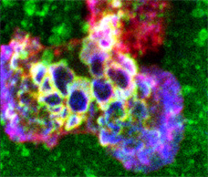


Supplemental Data to Podosomes Initiate Trans-cellular Diapedesis Immunity, 2007 ref. All Detailed Descriptions
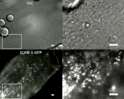
Video 1: Trans-cellular diapedesis is preceded by clusters of micron-scale ICAM-1-enriched rings on the endothelium.
Detailed Description
TNF-α-activated HDMVEC transiently transfected with ICAM-1-GFP were subjected to dynamic live-cell imaging upon addition of lymphocytes. Right panels correspond to the boxed regions in the left panels. Upper panels are DIC, lower panels are endothelial ICAM-1-GFP fluorescence. Note the dynamic ring-shaped ICAM-1-GFP-enriched structures are visible just prior to, and at the location of, trans-cellular pore formation. As diapedesis proceeds the trans-cellular pore expands to a maximum of ~ 6 μm before contracting and apparently resealing. The previously described (Carman and Springer, 2004) trans-migratory cup (comprised of vertical microvilli-like ICAM-1 projections that surround the trans-migration event) is transiently evident during diapedesis, though not ideally imaged at the selected focal plane. Several transient ring-shaped structures also form under a second lymphocyte. Video is a 70-fold acceleration of real time. Scale bars = 5 μm.
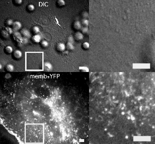
Video 2: Trans-cellular diapedesis is preceded by clusters of micron-scale ring-shaped perturbations the endothelium membrane topology.
Detailed Description
TNF-α-activated HDMVEC transiently transfected with memb-YFP were subjected to dynamic live-cell imaging upon addition of lymphocytes. Left panels correspond to a wide field of view in which most of a single GFP positive endothelial cell (in the context of a confluent endothelial monolayer) and multiple migrating lymphocytes are present. Right panels correspond to the boxed regions in the left panel and depict the initial stages of a lymphocyte trans-cellular diapedesis event. Upper panels show DIC and lower panels show endothelial memb-YFP fluorescence. Visualization of the endothelial cell surface with memb-YFP reveals dynamic ring-shaped structures formed under migrating lymphocytes. Rings precede formation of trans-cellular pores. Video is a 55-fold acceleration of real time. Scale bars = 5 μm.
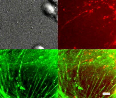
Video 3: Ring-shaped endothelial membrane structures form predominantly under the leading portion of laterally migrating lymphocytes.
Detailed Description
TNF-α-activated HLMVEC transiently transfected with memb-YFP (green) were subjected to dynamic live-cell imaging upon addition of lymphocytes. Upper panel is DIC (pseudo colored red). Middle panel is memb-YFP signal (green). Lower panel is the merge of the DIC and memb-YFP. A single lymphocyte migrates laterally from right to left a distance of ~35 μm and then initiates trans-cellular diapedesis. During the lateral migration, micron-scale endothelial transient ring-structures form predominantly in the region just behind the leading edge of the lymphocyte. Trans-cellular pore formation is initiated in the center of an expanding ring. Note also that initially the lymphocyte migrates over the endothelial cell nucleus (~15 μm diameter circular structure on the right side of the DIC image) forming ring-structures (albeit largely out of focus due to the raised topology of this region) directly on top of this organelle. Video is a 75-fold acceleration of real time. Scale bar = 5 μm.
Video 4: Endothelial cytoplasmic GFP is displaced at sites of ring formation.
Detailed Description
Video depicts initiation of a lymphocyte trans-cellular diapedesis event. Upper left panel is DIC. Upper right panel is memb-RFP fluorescence. Lower left panel is cytoplasmic GFP fluorescence. Lower right panel is a merge of the memb-RFP and GFP channels. Note that in the lower panels circular regions devoid of cytoplasmic GFP form at the location of each memb-RFP ring structure. Also note that the trans-cellular pore forms in the center of an expanding memb-RFP ring. Video is a 140-fold acceleration of real-time. Scale bar = 5 μm.
Video 5: Endothelial ER is displaced and distorted at sites of ring formation.
Detailed Description
Video depicts initiation of lymphocyte trans-cellular diapedesis event. Upper left panel is DIC. Upper right panel is memb-RFP fluorescence. Lower left panel is ER-Tracker Green fluorescence. Lower right panel a merge of the memb-RFP and ER-Tracker Green channels. Note that memb-RFP ring structures seem to form within the individual reticula of the ER often distorting and expanding these structures. Furthermore close examination of the rings demonstrates that irregular, sometimes trapezoidal, shapes that seem to correspond to the preexisting geometry of the ER reticula. Finally, note that the trans-cellular pore forms at the location of a membrane ring such that a ring present in one video frame is replaced in the next by the trans-cellular pore. Video is a 105-fold acceleration of real-time Scale bar = 5 μm.
Video 6:Endothelial actin-microfilament are displaced and distorted at sites of ring formation.
Detailed Description
Video depicts formation of endothelial ring structures on TNF-α-activated HLMVEC under adherent and laterally migrating lymphocytes. Upper left panel is DIC. Upper right panel is memb-RFP fluorescence. Lower left panel is GFP-actin fluorescence. Lower right panel is a merge of the memb-RFP and GFP-actin channels. Note that, in general, GFP-actin is displaced and in particular actin stress fibers are transiently distorted or “bowed-out” at sites of memb-RFP ring formation. Video is a 140-fold acceleration of real-time. Scale bar = 5 μm.
Video 7: Endothelial micro-tubules are displaced and distorted at sites of ring formation.
Detailed Description
Video depicts formation of endothelial ring structures on TNF-α-activated HDMVEC under a migrating lymphocyte. Upper left panel is DIC. Upper right panel is memb-RFP fluorescence. Lower left panel is GFP-tubulin fluorescence. Lower right panel a merge of the memb-RFP and GFP-tubulin channels. Note that, in general, GFP-tubulin signal is displaced and in particular that microtubules are transiently distorted or “bowed-out” at sites of memb-RFP ring structure formation. Video is a 150-fold acceleration of real-time. Scale bar = 5 μm.
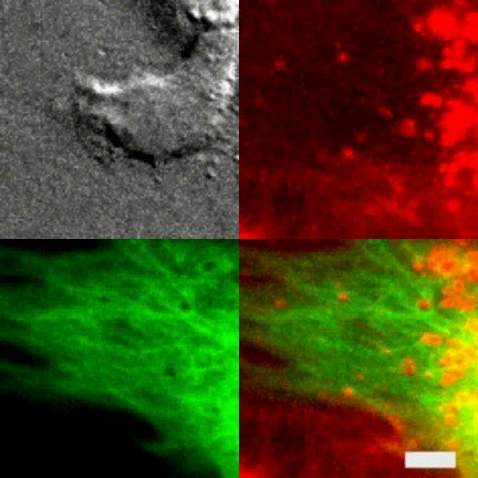
Video 8: Ring-structures form over the endothelial nucleus.
Detailed Description
Video depicts formation of endothelial ring structures on TNF-α-activated HLMVEC under a migrating lymphocyte. Upper left panel is DIC. Upper right panel is memb-RFP fluorescence. Lower left panel is cytoplasmic GFP fluorescence. Lower right panel is a merge of the memb-RFP and cytoplasmic GFP channels. Note, that GFP signal is displaced at sites of memb-RFP ring formation. As the lymphocyte passes over the nucleus (readily apparent in DIC, GFP and Merge panels and indicated with a white circle in frame 1), memb-RFP rings and displace GFP can be seen to from directly over the nucleus. Video is a 130-fold acceleration of real-time. Scale bar = 5 μm.
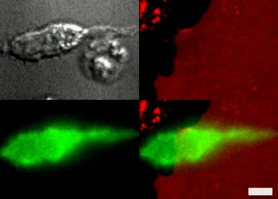
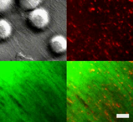
Video 9: Lymphocytes form dynamic podosome-like actin puncta at sites of endothelial invaginations.
Detailed Description
Video depicts endothelial podo-print induction on TNF-α-activated HDMVEC expressing memb-RFP under the uropod of lymphocyte expressing actin-GFP. Upper left panel is DIC. Upper left panel is memb-RFP fluorescence. Lower left panel is lymphocyte actin-GFP. Lower right panel is the merge of the memb-RFP and actin-GFP channels. Note the dynamic appearance of podosome-like actin puncta in lymphocytes that are spatially and temporally correlated with the formation endothelial podo-prints. Video is a 60-fold acceleration of real-time. Scale bar = 5 μm.
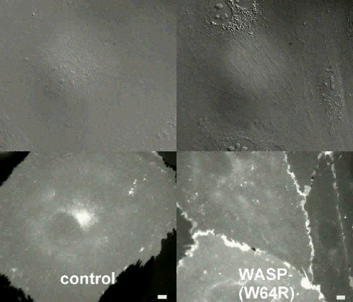
Video 10: Control and WASP-deficient lymphocyte migration and diapedesis.
Detailed Description
Video depicts a representative live-cell imaging experiment of control (left panels) and completely WASP deficient (W64R) (right panels) IL-2-cultured lymphocytes migrating on TNF-α-activated, memb-YFP-transfected HLMVEC. Upper panels show DIC. Lower panels show memb-YFP fluorescence. Though lymphocytes locally settled at different densities in the pre-selected fields, equal amounts of control and W64R lymphocytes were added. Note polarization, lateral migration and para-cellular diapedesis of W64R cells is generally similar to wild type cells, whereas podo-prints and trans-cellular diapedesis associated with these cell occurs relatively infrequently. Video represents a 100-fold acceleration of real time. Scale bars = 5 μm.
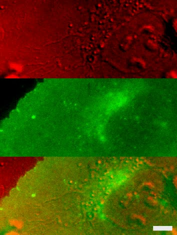
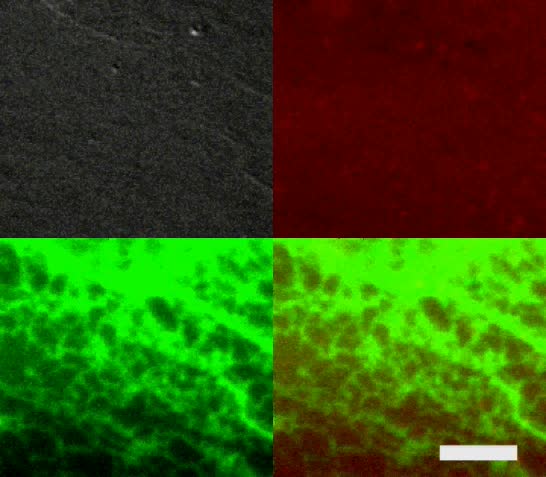
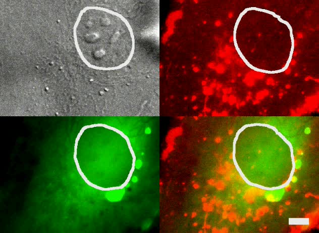
Video 11: Distribution dynamics of endothelial caveolin-1 with respect to podo-print and trans-cellular pore formation.
Detailed Description
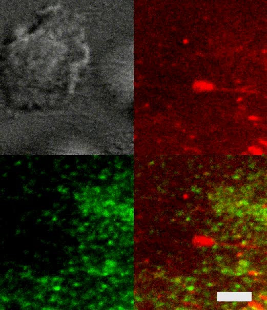
Video depicts representative live-cell imaging of podo-print and trans-cellular pore formation on TNF-α-activated HDMVEC co-expressing memb-RFP and caveolin-1-GFP. Upper left panel is DIC. Upper right panel is memb-RFP fluorescence (red). Lower left panel is cav-1-GFP fluorescence (green). Lower right panels are the merge of the red and green fluorescence channels. Note that on average low correlation exists between memb-RFP and cav-1-GFP signals both prior to and during podo-print formation under several migrating lymphocytes. A similarly modest correlation exists during initial formation of the two trans-cellular pores depicted. It is not clear whether the mild increase of cav-1-GFP around the perimeter of the lower trans-cellular pore during later stages of diapedesis represents an actual enrichment or simply the peripheral accumulation of the laterally displaced caveolin-1 as the pore expands. Video represents a 97.5-fold acceleration of real time. Scale bar = 5 μm.
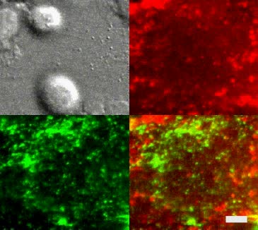
Video 12: Endothelial VAMP3 is enriched at podo-prints and initial trans-cellular pores.
Detailed Description
Video depicts representative live-cell imaging of podo-print and trans-cellular pore formation on TNF-α-activated HLMVEC co-expressing memb-RFP and VAMP3-GFP. Upper left panel is DIC. Upper right panel is memb-RFP fluorescence (red). Lower left panel is VAMP3-GFP fluorescence (green). Lower right panels are the merge of the red and green fluorescence channels. Note that whereas low correlation exists initially between memb-RFP and VAMP3-GFP signals, VAMP3-GFP develops significant local enrichment that is spatially and temporally correlated with memb-RFP signals in podo-prints and initial trans-cellular pore formation. Video represents a 90-fold acceleration of real time. Scale bar = 5 μm.
“Video for Everybody” v0.3 by Kroc Camen camen design